Products Who we are Employment Contact Us Home
 
Image Gallery
Optical Devices
Software
Confocal Equipment
Select Sample Gallery
hair

Biology
Brightfield

Leaf
Pancreas

Biology
Fluorescence

Bovine
Integrated Circuit

Industrial
Circuits

Integrated Circuit
Sandpaper

Industrial
Miscellaneous

Hexagon Pits
Microlens

SurfaceScope™ 3D Modeling

Micro-chip

-Image Acquisition-         -Image Types-


Image Acquisition:
The images were acquired with a standard Olympus BX50 microscope integrated with SufaceScope™ hardware. (Several other microscopes are supported including models from Leica, Nikon, and Zeiss.) No special sample preparation was involved; the samples were simply placed on the stage and illuminated with a standard halogen lamp.

SurfaceScope™ captures only the in-focus image data, disregarding all out-of-focus data. A focus controller was used to capture incremental focal planes, called "optical sections", at depths as fine as 0.1µ. The sections were projected to a computer by way of a mounted digital camera to allow for post acquisition processing with Image-Pro PlusŪ v4.1 and SurfaceView™ v1.0.

Image Types:
The SurfaceScope™ confocal system creates several different types of images (extended-focus composite, depth-colored, contour enhanced, and SurfaceScope™.

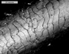
Extended-focus composite (EFC) images are based on optical sections of only in-focus data. This is the standard confocal mode in which all the in-focus optical sections are combined so that there is no out-of-focus data. (The hair fiber image above 'Biological Brightfield' is of this type.)

Depth-colored images take the EFC image a step further by adding color when recombining the optical sections. This method provides additional Z-dimension information through color. Generally the highest features are colored red down in rainbow fashion to the lowest which are colored blue. (The leaf image below 'Biological Brightfield' is of this type.)

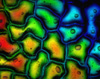
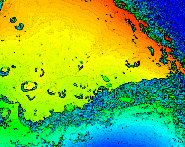
Contour enhanced images are an alternative to depth-colored. This method provides greater depth information by way of a caricaturization of the data. The colors are more separated to make finer depth judgements visually. (The sandpaper image above 'Industrial Miscellaneous' is of this type.)

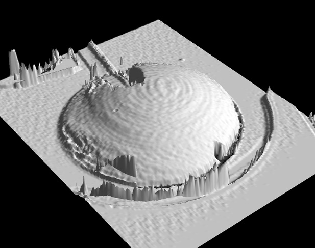
SurfaceScope™ images are created with the SurfaceView™ 3D modeling software. The images presented here are captures from the interactive software which allows rotation, resizing, and tilting. (This image type is present in the SurfaceScope™ gallery.)

Biology Brightfield
Biology Fluorescence
Industrial Circuits
Industrial Miscellaneous
SurfaceScope™ 3D Modeling