|
Chipped Auto Paint with Clearcoat
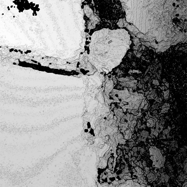
Figure 1 - Contour Enhancement: The topography is enhanced in this image taken at 100x magnification under standard halogen lighting.
Depth has been quantized and assigned shades of grey for more precise depth analysis. This paint chip image gives a dramatic impression of depth.
(Scroll for additional images of this sample.)
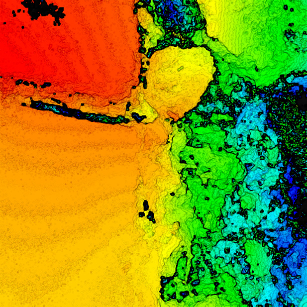
Figure 2 - Contour Enhancement: The topography above is further enhanced with the addition of color. Depth has been quantized and assigned unique colors for more precise
depth analysis. (Scroll for additional images of this sample.)
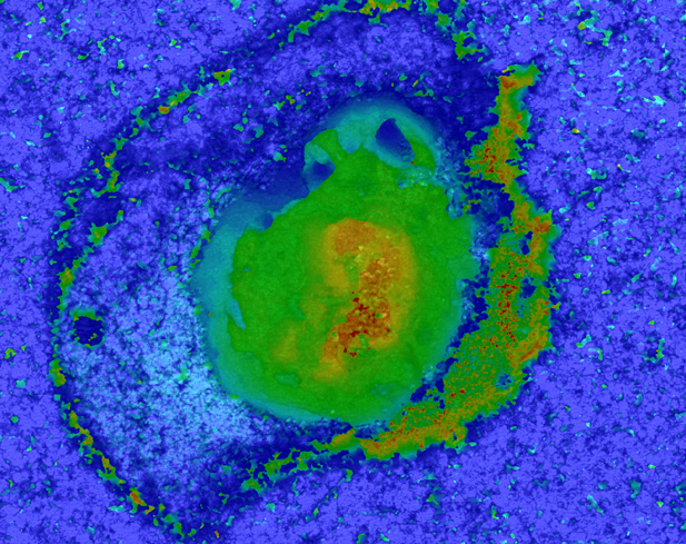
Figure 3 - Depth Colored: Depth information has been added to this image taken at 10x magnification under standard halogen lighting through color with blue corresponding
to the higher points and red the lower (this particular image has
the colors reversed relative to most images on this sight). This image reveals the cracking of the clearcoat on the edges and a sloped chipping toward the exposed metal surface (red) at the center.
(Scroll for additional images of this sample.)
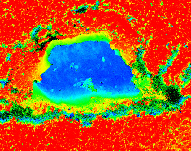
Figure 4 - Depth Colored: Depth information has been added to this image taken at 10x magnification under standard halogen lighting through color with red corresponding
to the higher points and blue the lower. This image reveals similar cracking
as above of the clearcoat on the edges and a sloped chipping toward the exposed metal surface (blue) at the center.
(Scroll for additional images of this sample.)
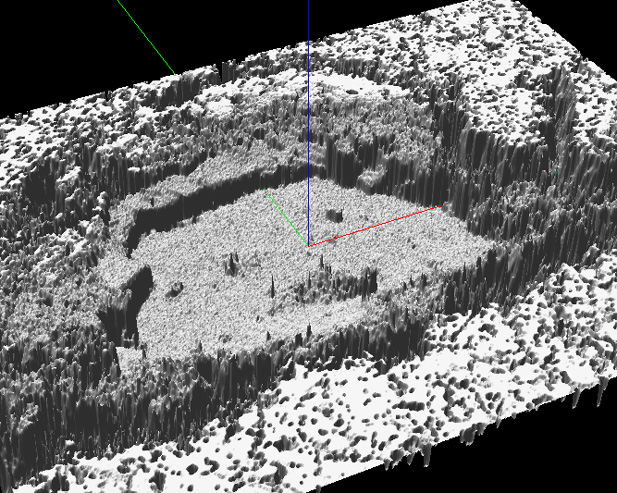
Figure 5 - SurfaceScope Capture: This 3D model of the above dataset allows rotation and tilting of the sample. (Scroll for additional images of this sample.)
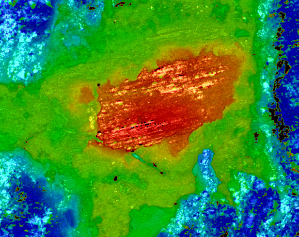
Figure 6 - Depth Colored: Depth information has been added to this image taken at 10x magnification under standard halogen lighting through color with red corresponding to the higher points and blue the lower(this particular image has
the colors reversed relative to most images on this sight). This image reveals a sloped chipping toward the exposed metal surface (red) at the center.
(Scroll for additional images of this sample.)
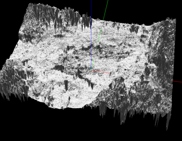
Figure 7 - SurfaceScope Capture: This 3D model of the above dataset allows rotation and tilting of the sample. (Scroll for additional images of this sample.)
| 