|
Fly Eye (Ommatidia)
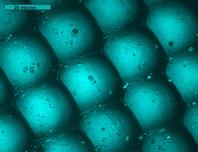
Figure 1 - Full-Focus Composite: This fly, found on our window sill, was imaged with a 100x air objective under standard halogen lighting. Dust particles are visible
throughout as are the small ridges between the ommatidium. (Scroll for additional images of this sample.)
.jpg)
Figure 2 - Depth Colored: Depth information has been added to the image above through color with red corresponding to the higher points and blue the lower.
The color reveals that this sample was at a rather sharp angle - each row of ommatidia are similarly colored as the depth covered a large range.
(Scroll for additional images of this sample.)
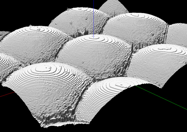
Figure 3 - SurfaceScope Capture: This 3D model allows rotation and tilting of the sample. (Scroll for additional images of this sample.)
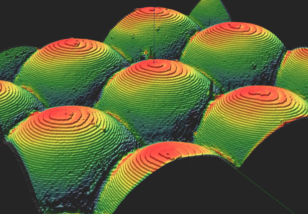
Figure 4 - SurfaceScope Capture: This 3D model allows rotation and tilting of the sample. (Scroll for additional images of this sample.)
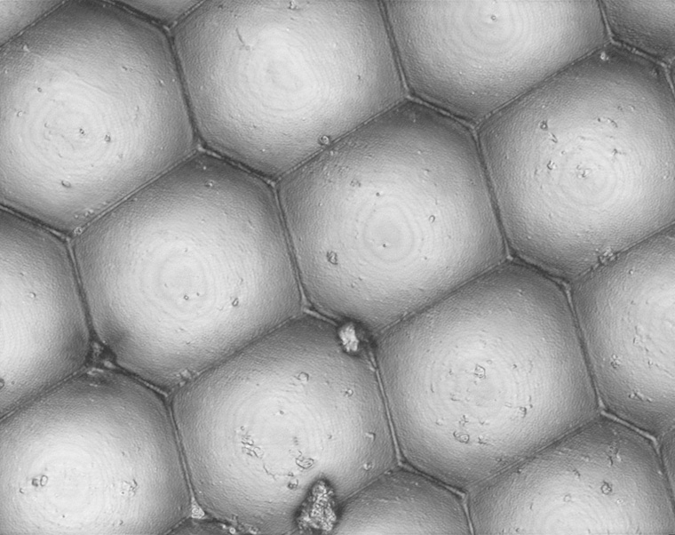
Figure 5 - Full-Focus Composite: This is another image from the same fly under the same conditions as above (100x), but along a flatter portion of the eye.
(Scroll for additional images of this sample.)
.jpg)
Figure 6 - Depth Colored: Depth information has been added to the image above through color with red corresponding to the higher points and blue the lower.
The color reveals that this sample was at a more shallow angle than above - the reduced depth range allows each ommatidium to include considerable depth information.
(Scroll for additional images of this sample.)
| 